You’ll Want To Know About This
Glaucoma is an eye disease that can lead to permanent vision loss in one or both eyes. The ailment often comes with no symptoms, remaining unnoticed until damage is done. Because of this, regular visits to an ophthalmologist are essential for catching glaucoma early and preventing ocular issues.
The risks aren’t worth it
No one is safe from glaucoma, but specific groups of people are more susceptible than others. Individuals are six times more likely to obtain the disease after age 60. This risk increases if there’s any family history of glaucoma. Furthermore, certain races such as African Americans, Asians, and Hispanics are more likely to have the ailment. Conditions such as diabetes, high blood pressure, and farsightedness can also play a role.
Test me; I dare you
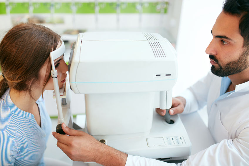
Since glaucoma is not always easy to diagnose, a series of tests have been created to locate underlying factors. Upon visiting the eye clinic, a physician may perform any number of these tests to check for signs of glaucoma. Each test checks a different aspect of the eye for indicators that glaucoma may be present. Patients with a higher risk for the disease may receive the whole gamut of tests at each exam. For others, some tests may only be necessary on an occasional basis.
Tonometry
Tonometry is a test that measures the pressure inside the eye. Most offices will numb the eye with drops and use a tiny instrument to touch the surface. The machine then records the amount of pressure coming from the eye. Since glaucoma causes damage by putting pressure on the optic nerve, a high number is an immediate red flag. For many, a pressure above 21mmHg is considered higher than normal.
Ophthalmoscopy
An ophthalmologist will typically use dilating drops to see more clearly into the back of the eye. Using a light and magnifier, the doctor is able to have a clear view of the optic nerve and retina. A careful study of the optic nerve can reveal damage caused by glaucoma or another condition that warrants attention.
Gonioscopy
The eye constantly recycles fluid through a drainage system located between the iris and cornea. If these channels become blocked, too much liquid remains in the eye, causing pressure to rise. A physician uses a special lens to check this area for obstructions that may be a precursor to glaucoma formation.
Pachymetry
The thickness of the cornea, or surface of the eye, can help interpret eye pressure readings. First, the eye is numbed using drops. A doctor or technician then places a small probe called a pachymeter on the cornea for an accurate measurement.
Perimetry
Also known as a visual field test, perimetry creates a map of the eye’s entire field of vision. Glaucoma usually consumes the outer edges of sight first, and this test can determine if any loss has occurred. During the test, patients will be asked to look straight ahead at a screen. Lights will appear on the sides of vision, and individuals must let the doctor know every time one is visible.
The meaning behind it all
Any abnormalities during these tests could be evidence of an issue brought upon by glaucoma or another ocular problem. While a glaucoma diagnosis is not great news, there are treatment options to relieve pressure and battle vision loss. In any case, catching the disease before problems occur offers the best chance of maintaining clear vision down the road.